Rannvá Dahl vart ph.d. á Københavns Universitet
Týsdagin 16. apríl vardi Rannvá Dahl ph.d.-ritgerð sína á Københavns Universitet. Ritgerðin kallast “Insulin stimulation of L-carnitine uptake in skeletal muscle in patients with primary carnitine deficiency and the metabolic state of the skeletal muscle”.
Rannvá Dahl hevur verið innskrivað til ph.d.-lestur á Biomedicinsk Institut á Københavns Universitet. Vegleiðarar hava verið Steen Larsen og Clara Prats Gavalda, bæði lektarar á Biomedicinsk Institut.
Granskingarráðið hevur stuðlað verkætlanini.
Í metingarnevndini vóru
Charlotte Mehlin Sørensen, lektari á Biomedicinsk Institut, Københavns Universitet, forkvinna, Ryan Godsk Larsen, lektari á Institut for Medicin og Sundhedsteknologi á Aalborg Universitet og Francis Stephens, professari á University of Exeter.
Verjan gekk sera væl.
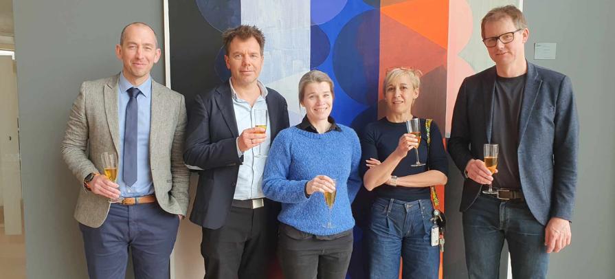
Á myndini er Rannvá Dahl í miðjuni saman við vegleiðarum og metingarnevndarlimum. © Privatmynd.
Enskur samandráttur
Background: Primary carnitine deficiency (PCD) is an autosomal recessive disorder characterized by a lack of functional carnitine transporters OCTN2 (Organic Cation/Carnitine Transporter 2), which has been linked to several cases of sudden death in young Faroese individuals. It causes low carnitine levels and can present with cardiac complications as well as skeletal muscle symptoms including muscular weakness and fatigue and studies have reported reduced fatty acid oxidation in relation to reduced carnitine content. Patients are treated with L-carnitine, and even while receiving treatment, skeletal muscle carnitine levels are only 5-20 %. The regulation of the carnitine transporter, OCTN2 is not fully understood, but a combination of hypercarnitinaemia with hyperinsulinemia upregulates skeletal muscle carnitine uptake and OCTN2 mRNA expression.
Objective: The question arises as to whether hyperinsulinemia increases the efficiency of L-carnitine supplementation in PCD. The primary aim was to investigate the regulatory mechanisms behind insulin-induced carnitine uptake, and whether the combination of hypercarnitinaemia and hyperinsulinaemia increases skeletal muscle carnitine levels in patients with PCD. Additionally, we wanted to examine if the low muscle carnitine has caused any mitochondrial and metabolic alterations in skeletal muscle of patients with PCD. Furthermore, we measured whole body measurements during exercise.
Method: Nine patients with PCD (homozygous for the c.95 A > G, P.N32S mutation), taking L-carnitine supplementation and nine healthy controls matched to age and body mass index (BMI) participated in the study (Study I, II). A six hour hyperinsulinemic clamp was supplemented with infusion of L-carnitine the last five hours. Skeletal muscle biopsies were collected before and after the clamp and carnitine content was measured. Mitochondrial function was measured as mitochondrial respiratory capacity and skeletal muscle H2O2 production in permeabilized muscle fibers. An attempt was done to measure mitochondrial respiratory capacity in intact fibroblast cell, (fibroblast-pilot study). Confocal microscopy was used to access regulation of OCTN2 positive vesicles due to insulin stimulation and to measure intramuscular lipid distribution. Additionally, indirect calorimetry was used to measure fatty acid oxidation at rest and during exercise.
Results and discussion: We found that the combination of hypercarnitinaemia with hyperinsulinemia did not increase skeletal muscle total carnitine levels significantly for neither patients with PCD (P = 0.28) nor controls (P = 0.053). Indicating that the attempt to increases the efficiency of L-carnitine supplementation in PCD did not succeed. The results from confocal microscopy suggested that insulin regulates skeletal muscle carnitine uptake by stimulating OCTN2 recruitment from intracellular storages to the plasma membrane in controls, a mechanism that seems to be impaired in patients with PCD.
Even though, the regulatory mechanism seems to be impaired there are other mechanisms associated with OCTN2 recruitment to plasma membrane, such as muscle contraction. Which current study did not investigated. Furthermore, we found the mitochondrial respiratory capacity, in permeabilized muscle fibers, to be similar between groups, as well as the H2O2 emission and intramuscular lipid distribution. Indicating that the reduced levels of muscle carnitine, reported in patients with PCD (taking L-carnitine) do not seem to have damaged the mitochondrial complexes of skeletal muscle and that even though carnitine has been found to have antioxidant properties, the levels of H2O2 were not increased in skeletal muscle of patients with PCD. The muscle free carnitine in PCD was low, as expected, however the acetylcarnitine levels were within the normal range, which could indicate that carnitine´s role as an acetyl buffer is intact. This buffering role is crucial during high-intensity exercise which aligns with the observation that patients with PCD demonstrated a VO2Max exercise test performance comparable to that of the control group. Additionally, their fatty acid oxidation during submaximal exercise was also similar to the control group.